Many injuries and ailments require medical imaging to help determine a diagnosis. Its common to have imaging scheduled to learn more about what is causing you pain. So what does each type of imaging show? Let's take a closer look into the different types of medical imaging.
X-Ray
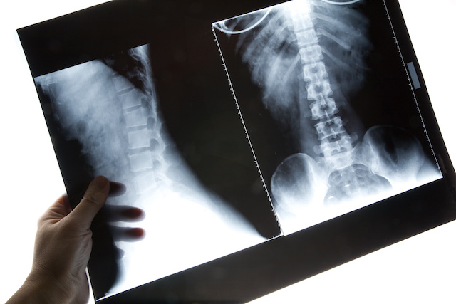
X-Rays shows imaging through x-ray beams passing through your body and absorbing in varying amounts depending on the density of what is passes through. Dense substances such as bones show up white, and air shows up black. Different densities of tissues show up in shades of gray.
Diagnosing problems in the bones and teeth are the most common uses for x-rays. The lungs, heart and breast tissue are also areas where MRI's are used to detect anomalies. In some cases, a contrast dye is used to better detect problems with the digestive track and blocked blood vessels.
MRI
Magnetic resonance imaging, or MRI, uses a strong magnet and radio waves to produce detailed images of the body by computer. Because MRI's use a magnetic field, the patient can not wear any metal during the imaging process. Patients with implanted medical devices or other foreign bodies containing metal are most likely unable to have an MRI. In some cases a dye is used to show contrast with the imaging, similar to x-rays.
MRI's are used for many reasons. Most commonly, they are used to find abnormalities in the spine and the tissues surrounding joints. Problems with the heart, brain, liver, breast, uterus, and abdominal organs can also be found with an MRI.
CAT Scan
Computed Axial Tomography, known as a CAT scan or CT scan, uses x-rays along with a computer to produce cross sectional images of the body. They are able to produce a mote detailed images than a normal x-ray machine by using thin slices of the body for each image.
CAT scans provide more details than x-rays, making them better to pinpoint fractures, tumors, blood clots, infections, and internal bleeding. Treatments for cancer can use CAT scans to help monitor effectiveness. They also can provide better mapping for surgical treatments and biopsies.
Bone Scan
Bone scans relate to just what the name implies- they provide imaging of the bones. A small amount of radioactive dye used during the procedure and will accumulate in the parts of the bone tissue that are undergoing a physical or chemical change. The radiation produced in these areas will show up on the scanner. The radiation used is small and is release from the body completely within a few days.
Bone cancers and cancer metastasizing to the bones are the primary use for bone scans. They also detect any type of weakness in the bones, which can identify arthritis, osteoarthritis, and infections not seen in other types of imaging.